1. Application Overview
3D printing (RP rapid prototyping) is to use stacking technology to output your three-dimensional image files into real 3D models, such as upper and lower jaw, hand bones, teeth, etc...
2. System Overview
The new office model ProJet HD3000 3D printer developed by 3D Systems is just up to this task. As long as you convert 2D tomographic 3D scans into 3D solid files, then the HD3000 3D printer will not only save manual work The time and cost of making the model make the patient no longer have to endure the pain of waiting
3D Printing 3D Printing Surgery Application

3. Application field
The current surgical reconstruction includes the treatment process of craniofacial plastic surgery, orthopedics, and dentistry. The doctor schedules treatment procedures based on the patient's 2D plane X-ray or tomographic data, and uses his professional and long-term accumulated experience to judge the condition. Since doctors get flat data, blind spots will inevitably occur in judgment, and patients cannot clearly understand the treatment situation. This not only increases the medical risks, but may also cause medical disputes, which is not worth the gain.
Therefore, we propose an integrated solution for medical treatment and 3D printing technology that will greatly improve the quality of medical treatment and is a breakthrough in medical technology. With these state-of-the-art and mature technologies, doctors will be able to actually hold a 3D three-dimensional model of the patient's skull and bones, make the most accurate judgments, and directly perform treatment scheduling and surgery simulations.
4. Application process
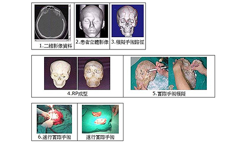
Application of 3D printed skull plastic surgery
5. Successful cases of medical and 3D printing: the application of cranial plastic surgery
The success of craniofacial plastic surgery not only directly or indirectly affects the patient's appearance, but also affects the symmetry and integrity of the skeleton that supports the overall appearance. With traditional plastic surgery, most surgeons perform operations based on their personal experience with two-dimensional computed tomography images. Due to the lack of three-dimensional image processing software and solid models to assist in the simulation before surgery, the key to the success of the surgery is the surgeon’s Clinical Experience. However, performing surgery based solely on the physician's personal experience and awareness often results in disagreement between the physician and the patient on the surgical method after the operation.
To improve the aforementioned bottleneck that clinicians lack stereoscopic images and solid models to assist in the simulation of surgery, we reorganized the patient's computed tomography images into 3D stereoscopic images on the computer in advance, and used 3D printing technology to generate 3D printed solid models, so that Before performing craniofacial plastic surgery, the surgeon can simulate the cutting displacement and spatial positioning of the stereo image on the computer, and use the solid model to plan the surgical path and analyze the situation that may occur during the operation. This can make the surgeon and the disease The patient’s family members can have a more detailed understanding of the entire surgical planning before the operation. In addition to reducing the communication difficulties and misunderstandings between the doctor and the patient, virtual images and physical models of the surgical planning and simulation can also be used to enable the doctor to practice in advance. It will improve the quality and success rate of the operation, and will positively help the patient's postoperative recovery and the safety of the operation.
![]() Tel:+86 135 7086 9158
Tel:+86 135 7086 9158![]() Tel:+86 135 7086 9158
Tel:+86 135 7086 9158
